| Plague in Southern Caucasus |
|
D. Tsereteli 1, L. Bakanidze
1, I. Velijanashvili 1, L. Beridze 1, M. Manrikyan 2, M. Shakhikyan
2, M. Kekelidze 1, E. Zangaladze 1, N. Tsertsvadze 1, P. Imnadze
1 |
| Key-words:
Plague, Yersinia pestis, PCR, natural foci |
Summary There are three natural foci of plague on the territory of Georgia and Armenia: plain–foothills, high-mountainous and Araks-side. High-mountainous focus includes the southern part of Georgia and the eastern part of Armenia. Conventionally by state border it is divided into two parts. Strains of Y. pestis isolated from high-mountainous focus differ from strains of Y. pestis isolated in plain–foothills and Araks-side foci. In this study 21 strains of Y. pestis, isolated from high-mountainous focus, 1 strain of Y. enterocolitica and 2 strains of Y. pseudotuberculosis were tested by PCR. All 21 strains of Y. pestis produced amplicons in the V antigen gene PCR. 19 of them produced amplicons in the F1 capsular antigen gene PCR, 2 strains were negative. All strains lacked pPLA antigen gene. |
Schlüsselwörter: Pest, Yersinia pestis, PCR, Naturherd |
Zusammenfassung Auf dem Territorium von Georgien und Armenien finden sich drei unterschiedliche Naturherde für Pest: Ebenen/Hügellandschaften, Hochgebirge und Gebiete am Fluss Arax. Hochgebirgsherde sind im südlichen Georgien und im östlichen Armenien platziert. Yersinia pestis-Isolate von Hochgebirgsherden unterscheiden sich klar von Isolaten, welche aus Naturherden in Ebenen/Hügellandschaften oder Gebieten am Fluss Arax stammen. Wir untersuchten 21 Hochgebirgs-Isolate von Yersinia pestis im Hinblick auf Virulenzgene. Yersinia enterocolitica (1 Isolat) und Yersinia pseudotuberculosis (2 Isolate) wurden als Kontrollstämme verwendet. Mittels PCR konnte bei allen Yersinia pestis-Isolaten das Gen für das V-Antigen nachgewiesen werden. Bei 19 dieser Isolate konnte Plasmid-Nukleinsäure nachgewiesen werden, die das F1-Kapselantigen codiert. Allen Isolaten fehlte das pPLA-Antigen; die Abwesenheit dieses Virulenzfaktors gilt als Besonderheit der Yersinia pestis-Isolate des südlichen Kaukasus. |
| Introduction
Yersinia pestis is one of the
most devastating infectious agents in the world. Cases of plague
were described starting from the pre-Christian era. Despite of major
advances made in the knowledge of the disease, plague has not been
eradicated yet. Today, in the perspective of the last events the
threat of bioterrorism made the society more sensitive to those
infectious agents, which can be used as a biological weapon. And
plague is one of the main threats of bioterrorism.
|
| Materials
and Methods
Materials for the investigation were
taken from live rodents and rodent remains from locations of epizootics:
parts of spleen, liver, lungs, pathologically changed lymph nodes,
bone marrow and brain. Fleas and ticks were combed out, and collected
in burrows. Suspected on plague, material was investigated by microscopy,
cultivation on elective media, serology and infecting laboratory
animals. Isolated cultures were investigated by bacteriological,
biochemical and biological methods. All the strains were analyzed
by PCR that was performed using ready-to-go analysis beads („Pharmacia
Biotech“). Bacterial DNA for PCR was derived by the standard
methodology [1]. The primer pairs used for identifying F1 capsular
antigen gene and for V antigen gene were as described [2]. PCR for
F1 capsular antigen gene was performed using the following cycle
profile: |
| Results
and Discussion
There are three natural foci of plague on the territory of Georgia and Armenia: plain–foothills, high-mountainous and Araks-side. Plain–foothills focus includes the eastern part of Georgia and Azerbaijan territories near the Azerbaijan-Georgia border. Epizootics of plague in the Georgian part of this focus were registered in 1966 and 1968-1971 [3, 4]. The main reservoir of plague in these foci is Meriones erythrourus (libicus), the main vector – Xenopsylla conformis and Ceratophyllus laeviceps. Totally 83 strains were isolated: 30 strains from rodents and 53 strains from the vectors. High-mountainous focus includes the southern part of Georgia and the eastern part of Armenia. Conventionally by state border it is divided into two parts. High-mountainous focus of southern Caucasus first was identified in 1958, when the Y. pestis strain was isolated from Microtus arvalis in the Giumri neighborhood. In the Armenian part of this focus every year in the period of 1958-1988 there were epizootics of plague of different intensity. It must be mentioned that after the earthquake in Armenia in 1988 and until the end of the 1990s epizootic processes decreased in the Armenian part of the focus, and reservoirs and vectors of plague were heavily depressed. Beginning from the year 2000, epizootic processes are activated. In the Georgian part of the focus epizootics of plague were identified in the years 1979-1983 and 1992-1997. The main reservoir in this focus was Microtus arvalis, the main vector – Callopsylla caspia, Nosopsyllus consimilis. From 39 strains isolated 5 strains were from rodents and 34 from vectors. In 1971 Araks-side focus of plague was identified. The main reservoir in this focus was Meriones vinogradovi, the main vector – Xenopsylla conformis and Callopsylla iranicus. Strains of Y. pestis isolated from high-mountainous focus differ from strains of Y. pestis isolated in plain–foothills and Araks-side foci by several characteristics: they ferment ramnose during 48 hours, are less virulent or avirulent for guinea pigs, do not produce pesticin, but are sensitive to it. Pathogenic Y. pestis-isolates are characterized by the presence of three different plasmids. The Y. pestis - specific 9,5 kbp plasmid pPla carries the plasminogen activator/coagulase and pesticin genes [2, 9]. The other Y. pestis-specific plasmid is pFra, 110 kbp in size. The highly immunogenic fraction F1-capsular antigen and the murine toxin are well-studied proteins encoded by this plasmid. The third plasmid is 70 kbp in weight and is shared by all pathogenic members of the species Y. pestis, Y. enterocolitica and Y. pseudotuberculosis. It contains a number of highly conserved virulence marker genes, and codes for the V antigen, a factor in Yersinia supposed to influence the inflammatory response [1, 8]. Only the 70 kb plasmid is shared among the pathogenic yersinia [5, 7]. In this study 21 strains of Y. pestis,
isolated from high-mountainous focus, 1 strain of Y. enterocolitica
and 2 strains of Y. pseudotuberculosis were tested by PCR.
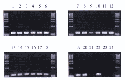
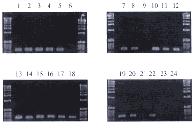
All 21 strains of Y. pestis produced amplicons in the V antigen
gene PCR (Fig. 2). 19 of them produced amplicons in the F1 capsular
antigen gene PCR, 2 strains were negative (Fig. 3). All strains
lacked pPLA antigen gene. This finding correlates with the data
that strains from Transcaucasian Alpines and Daghestan Mountains
are exceptions – they do not consist of 9,5 kbp pPLA [6]. 2
strains of Y. pseudotuberculosis and the strain of Y.
enterocolitica did not produce amplicons in any of three –
V, F1 and pPLA antigens gene PCR. Further studies of these strains
(PFGE) will show the correctness of these findings.
|
| References:
1. Brubaker B.: „The V antigen of Yersiniae: an overview.“ In: Une T., Maruyama T. and Tsubokura M. (eds), Current Investigations of the Microbiology of Yersiniae. Karger, Basel (1991) 127-133. 2. Campbell J., Lowe J., Walz S., Ezzell J.: „Rapid and specific identification of Yersinia pestis by using a nested polymerase chain reaction procedure.“ J. Clin. Microbiol. 31 (1993) 758-759. 3. Cherchenko I., Dzebisashvili Iu., Nersesov V., Oganyan E., Magradze G., Mchedlidze G., Velijanashvili I.: „Detection of the Specific Plague Antigen F1 in the Soil from the Tombs of the 19th Century in the Caucasus. Problems of Particularly Dangerous Infections (annals of the Plague Prevention Institutions of the USSR.“ Saratov, 6 (1973) 107-108. 4. Chkheidze G., Dzebisashvili Iu., Nersesov V., Jmukhadze I.: „Natural Foci of Plague on the Territory of Georgia. J. Particularly Dangerous Infections in Caucasus.“ Stavropol. 1 (1974) 70-72. 5. Chu M.C.: „Laboratory Manual of Plague Diagnostic Tests.“ CDC – WHO. (2000) 10-40. 6. Filippov A.A., Solodovnikov N.S., Kookleva L.M., Prostenko O.A.: „Plasmid Content in Yersinia pestis-strains of different origin.“ FEMS Microbiol. Lett. 67 (1990) 45-48. 7. Neubauer H., Meyer H., Prior J. et al.: „A Combination of Different Polymerase Chain Reaction (PCR) Assays for the Presumptive Identification of Yersinia Pestis.“ J. Vet. Med. B 47 (2000) 573-580. 8. Price S.B., Leung K.Y., Brave S.S., Straley S.C.: „Molecular analysis of lcrGVH, the V antigen operon of Yersinia pestis.“ J. Bacteriol. 171 (1989) 5646-5653. 9. Podzorsli R.P., Kukuruga D.L., Long P.M.: „Introduction to Molecular Methodology.“ Manual of Clinical Immunology, 5th Ed., Washington, DC; ASM Press (1997) 77-107. |
|
Corresponding author:
|


